
Experts in the fields of material analysis and lab-based testing, our team of highly trained specialists constantly strives to guarantee the safety and satisfaction of our customers, as well as the end-users and consumers of the products and services we collaborate on and contribute towards.
Owing to our strong company culture and commitment to the wider community, we believe that cooperation, not competition, between innovators, analysts, and regulators is integral to the supply of highly complex services and solutions.
Every plan and process we follow carries a strict protocol-based procedure to ensure maximum levels of safety through standardization and evaluation. Our approach to scanning electron microscopy (SEM) carries the same sense of pride and responsibility as any other research task we embark upon.
An Introduction to Scanning Electron Microscopy
Scanning electron microscopy is a technique used to produce high resolution (approximately 200x better than that of traditional optical microscopy) and high magnification images of a variety of samples by scanning in its entirety with a self-contained electron beam.
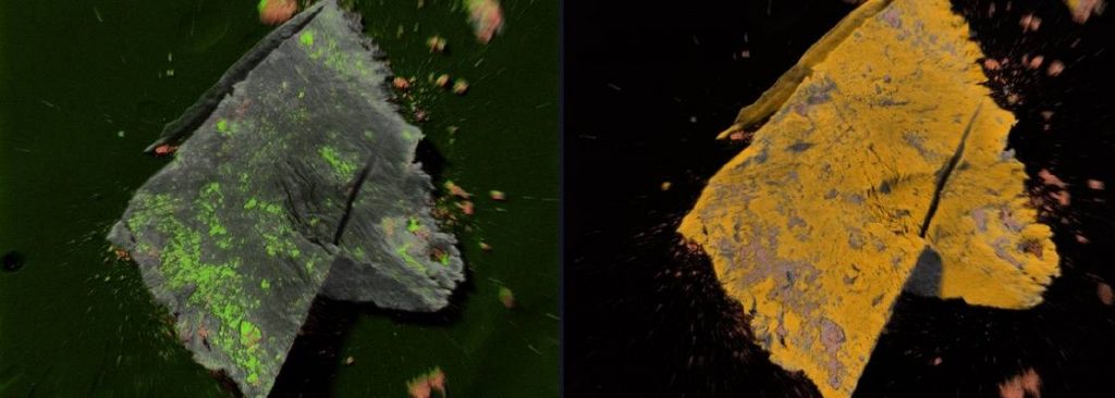
The interaction of the electron beam from the SEM and the specimen produces x-rays which can be detected by an energy dispersive x-ray spectrometer (EDS) to provide elemental composition.
The diversity and, in most cases, preservative powers of scanning electron microscopy analysis, allow researchers the luxury of a highly accurate reading without causing damage or deterioration to the original state of the sample.
The 3 Main Objectives of Scanning Electron Microscopy
1. To Generate High-Resolution and High-Magnification Images
Newfound techniques allow a more intrusive and insightful investigation of small-scale samples and physical surface structures (topography), giving more reliable readings down to tens of nanometers (nm).
2. To Depict a Clear Picture of Chemical Composition
Coupling SEM with EDS allows for the collection of high-resolution elemental maps. Mapping analyses allows for the visualization of elemental dispersion in samples – particularly useful for investigating API and excipient dispersion in drug products.
3. To measure particles and layers
Particles and layers can be measured down to nm sizes! The high-resolution nature of SEM allows for the measurement of layers and fine structure of matter previously impossible to see, read, and rate. The power to identify and analyze microscopic material provides us with the possibility to fully comprehend sample morphology.
Contact a member of the Jordi Labs team today to learn more about scanning electron microscopy and EDS detection.